A Legacy of Innovation and Family Dedication
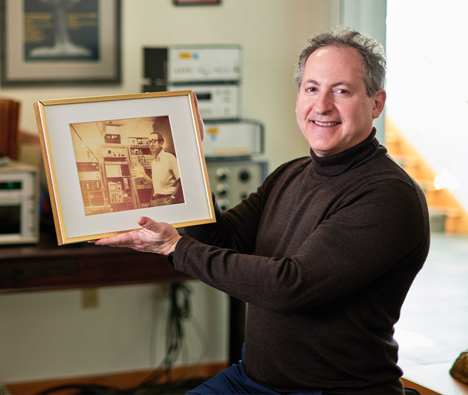
The origins of MBF Bioscience can be traced back to the visionary work of our co-founders, Jack Glaser, an innovative software developer, and his father, a pioneering neuroscientist and engineer whose groundbreaking research and commitment to innovative engineering laid the foundation for the company’s mission. What began as a family endeavor has evolved into a global enterprise, but our roots remain deeply intertwined with a lifelong dedication to advancing scientific knowledge and supporting researchers in their quest for new insights.
To learn more about the Glaser family’s remarkable journey and how their unwavering passion for science shaped the birth of MBF Bioscience, please explore our company history.