A method of mapping atlas information to a histological section image of a histological section
MBF Bioscience was just awarded a patent by the USPTO for our new brain mapping technology! This novel technology is now
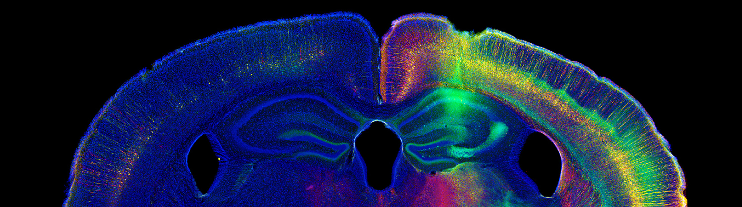

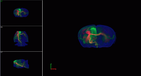
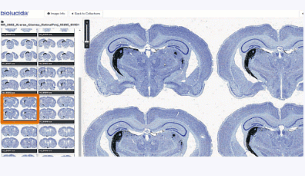


MBF Bioscience was just awarded a patent by the USPTO for our new brain mapping technology! This novel technology is now
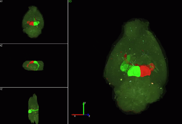
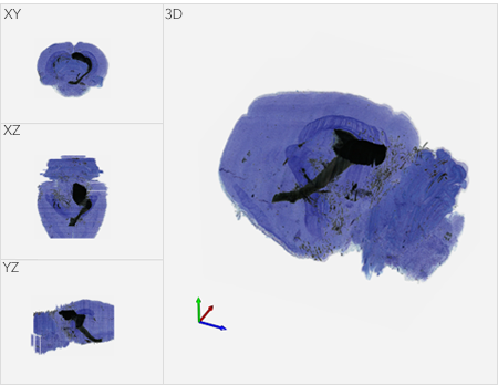
MBF Bioscience now offers customized models of Huron Digital Pathology’s TissueScope™, a line of whole slide scanners, and supports TissueScope images
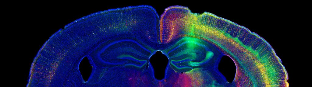
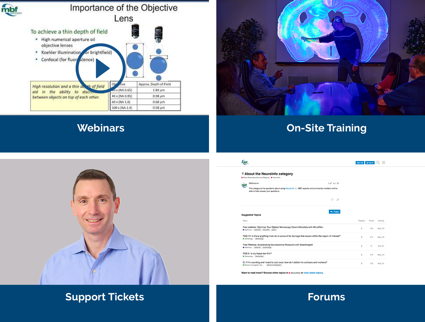
Analyzing cellular populations within specific anatomies in brain images requires expertise in both neuroanatomy and cellular identification. This typically involves
Gergues, M. M., K. J. Han, et al.
“Circuit and molecular architecture of a ventral hippocampal network.”View Publication

Lindberg, P. T., J. W. Mitchell, et al.
“Pituitary Adenylate Cyclase-Activating Peptide (PACAP)-Glutamate Co-transmission Drives Circadian Phase-Advancing Responses to Intrinsically Photosensitive Retinal Ganglion Cell Projections by Suprachiasmatic Nucleus.”View Publication

Paletzki, R. and C. R. Gerfen
“Whole Mouse Brain Image Reconstruction from Serial Coronal Sections Using FIJI (ImageJ).”View Publication

Zepecki, J. P., K. M. Snyder, et al.
“Regulation of human glioma cell migration, tumor growth, and stemness gene expression using a Lck targeted inhibitor.”View Publication

Zhang, L., V. S. Hernandez, et al.
"Behavioral role of PACAP signaling reflects its selective distribution in glutamatergic and GABAergic neuronal subpopulations." eLife 10: e61718.View Publication

Weber-Adrian, D., R. H. Kofoed, et al.
“Systemic AAV6-synapsin-GFP administration results in lower liver biodistribution, compared to AAV1&2 and AAV9, with neuronal expression following ultrasound-mediated brain delivery.”View Publication

"I rarely have encountered a company so committed to support and troubleshooting as MBF."

"MBF Bioscience is extremely responsive to the needs of scientists and is genuinely interested in helping all of us in science do the best job we can."

"I am so happy to be a customer of your company. I always get great help related with your product or not. With the experienced members, you are the best team I've ever met. All of your staff are very kind and helpful. Thank you for your great help and support all the time."

"We’ve been very happy for many years with MBF products and the course of upgrades and improvements. Your service department is outstanding. I have gotten great help from the staff with the software and hardware."

"Our experience with the MBF equipment and especially the MBF people has been outstanding. I cannot speak any higher about their professionalism and attention for our needs."
"MBF provides excellent technical support and helps you to find the best technical tools for your research challenges on morphometry."